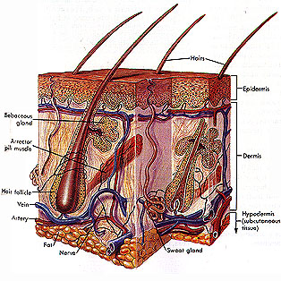
The Skin
The following information is regarding the
structure and function of the skin, including the appendage structures
derived from the skin (hairs and nails) and structures below the
surface of the skin (glands and sensory receptors). You will also
learn about some of the common diseases of the skin and way the
skin changes over the normal life span from infancy to old age.
The skin is a complex organ that includes different
tissues and structures whose origins lie within other systems.
Thus its activities must be coordinated with those of other systems,
espcially the circulatory, nervous, and endocrine systems. This
coordination is refered to as integrated functioning, and it enable
the body to maintain a stable internal environment for the healthy
operation of all cells and systems, even when conditions outside
or inside change.
This maintenance of stable internal conditions
is called homeostasis. Homeostasis is a concept that is essential
to understanding the functioning of the human body.
The Skin: An Overview
As the body's outermost covering, the skin
interacts directly with the environment and has several important
functions. Amongst these diverse functions are limits on the entry
and exit of materials, providing sensory awareness of our surroundings,
and the ablity to repair itself upon injury. Clearly the skin
is not merely a passive wall around the body.
- Protection and defense. The skin is an effective barrier against mechanical
injury and the absorption of dangerous chemicals. It blocks penetration
by all but sharp objects. It is the first line of defense against
foreign agents, such as bacteria and viruses, which cause disease.
- Sensation.
The skin is an early warning system for the body. Specialized
sensory organs in the skin continuously monitor the sensations
of touch, pressure, pain, warmth, and cold.
- Water balance.
The outermost layer of the skin contains a water-resistant protein
called keratin. This waterproofing reduces the loss of the body's
precious internal water.
- Temperature regulation and excretion of
wastes. In humans, the skin plays
a direct role in limiting heat loss if the body is too cool and
cooling the body if it becomes too warm. To achieve cooling,
sweat glands secrete water onto the skin. As it evaporates, this
water carries with it heat energy, cools the skin, and lowers
body temperature. The sweat glands also secrete salts and small
amounts of waste products such as urea and ammonia.
- Synthesis of vitamin D. Exposure to the ultraviolet wavelengths of sunlight
causes the skin to produce small quantities of vitamin D which
supplement the smount supplied in the diet. This vitamin is essential
to the contruction of bones and teeth from the minerals calcium
and phosphate.
Tissue
Structure of the Skin
 |
Functions of the skin as they were described
above are accomplished by two relatively thin layers of tissue:
an outer epidermis and inner dermis. These are
connected by a thin basement membrane which is somewhat anchored
into the dermis. The thinner epidermis is constructed from stratified
squamous epithelial tissue, while the thicker dermis is a
dense connective tissue. The skin ranges in thickness
from 0.5 mm over the eyelids to 6mm (about 1/4 in.) or more on
areas of the hands and feet that recieve heavy wear and tear.
|
The Epidermis:
a Thin Outer layer.
| The epidermis consists up to
five different layers, or strata. Of the five we will examine
only two layers: the innermost layer called the stratum basale
and the outermost layer, called the stratum corneum. The
epidermis varies in thickness, and all five strata are present
only in thick-skinned areas on the palms and the soles of the
feet. The epidermis constantly undergoes growth and renewal.
In regions of repeated pressure and friction, production of new
cells is stimulated. The skin will formed a callus or corn as
a result of pressure from abnormal wearing, so as you know, the
epidermis can form thick layers to protect the underlying dermis
from abrasion. |
 |
New cells are produced by mitoic cell
division in the basal stratum. These
cells are somewhat columnar and upon division, one daughter cell
remains in the basal region, the other migrates towards the surface.
This may take up to 56 days. As the new cells are produced, the
older cells are pushed upward toward the outer surface of the
skin, the cornified stratum.
As these cells move upward, they undergo distinctive
changes, the most significant of which is the synthesis of massive
amounts of the protein keratin, which eventually fills
the cells.The cells that make keratin are called keratinocytes
and account for about 95 percent of all the cells in the epidermis.
Due to keratinization, change shape and eventually die. These
dead squamous cells form the stratum corneum, a layer made of
as many as 25 layers or more. In addition to waterproofing, keratin
contributes great strength and toughnes to the skin and its appendage
structures, the nails and the hair.
The color of the skin, as seen by the appearence
of the outer layer of cells is determined by the thickness of
the stratum corneum, the underlying blood vessels, and the amount
of the brown-black pigment melanin. Melanocytes are a second
type of cell found in the epidermis (stratum basalae). They are
specialized to produce a dark pigment called melanin. These
cells produce and then pass on to other surrounding cells, throughthin
cytoplasmic extensions, the pigment. This process is refered to
as cytocrine secretion. Although regions of the body may
vary in degree of pigmentation, all people have approximatly the
same number of melanocytes, despite racial variation in color.
The amount of melanin produced is determined
by genetic factors, hormones, and exposure to light. Albinism
is a single genetic defect (even though many genes effect pigmentation
production) which prevents pigment production. Hormaonal changes
during pregnancy can lead to a greater amount of pigmentation
to be produced. Increased or decreased blood flow can change the
'pinkness' of the skin, and a decrease in oxygen content can lead
to a blueing of the skin (cyanosis). Carotene is
a yellowis-orange pigment which is lipid soluble and can be stored
in the lipds of the skin. Excessive intake can give the skin an
overly yellowish tint.
Melanin protects the DNA of the dividing cells
in the basal stratum from damage by ultraviolet wavelegnths of
sunlight. Changes in basal cell DNA can lead to skin cancer, and
this melanin screen provides some protection. Skin cancer can
be a serious consequence of extensive exposure to sunlight.
As with all epithelial tissues, the epidermis
contains no blood vessels. Cells in the lower strata can obtain
oxygen and nutriants only by diffusion from the dermis, which
is well supplied with blood vessels. As the cells reach the upper
strata, however, diffusion of nutriants is diminished and the
cells die.
The surface of the skin we admire and care
for consists of dead cells.Each day millions of dead skin cells
are flaked away, to be replaced with new cells from below. Normally
it takes about 4 to 6 weeks for cells to move from the basal stratum
to the cornified stratum, where they are lost. Some estimates
suggest that 80 percent of dust in our homes is actually dead
epidermal cells. Excessive flaking of cells produces dandruff.
The Dermis: the
Deep, Thick layer
The dermis thicker inner layer. The
dermis lies below the epidermis and is substantially thicker than
the epidermis. It is a layer of connective tissue that consists
of an extracellular matrix with abundant collagen, elastic, and
reticular fibers.
The dermis is made of two basic layers: the
upper papillary layer and the deeper reticular layer.
The reticular layer is primarily collagen and elastic fibers and
is responsible for providing the skin with it's strength. Although
collagen fibers lie in a multitude of directions within this layer,
there is a more specific direction that they may lie, dependent
on location. This allows the skin to have a greater flexibility
in directions of common 'stretch'. This orientation causes what
are commonly called cleavage lines. Damage across these lines
such as with excessive stretching or an incision of some sort)
will likely scar; damage with the line will produce little if
any marking.
The papillary region is called such by a series
of projections, papillae, that stretch upward into the
epidermis. These help anchor the demis into the epidermis and
are most numerous in regions with the greatest amount of wear:
the hands and feet. Within the hands and feet, these ridges form
elaborate parallel patterns, increasing friction, providing improved
gripping power. The papillary region also contains numerous blood
vessels to provide the epidermis with nutrients, remove waste,
and help regulate body temperature.
The hair follicle is also found within the
dermis, as are various glands, smooth muscle cells, sensory receptors
and their associated nerves. Vitamin D production occurs in the
dermis, stimulated by the sunlight which passes through the melanin
protection of the epidermis.
The Hypodermis:
the Underlying layer
Beneath the dermis is a layer called Hypodermis
or the Subcutaneous layer. While this is not considered
part of the integumentary system, it helps support the two layers
of the skin by anchoring them to the muscle or bone below. This
layer is made of loose connective tissue, including approximatly
half the bodies fat (adipose), depending upon gender, age,
and heredity. The fibers of the connective tissue is basically
continous with the dermis, so no real, defined boundry exists
between the two. This fat serves as padding for delicate underlying
structures and insulation for retaining body heat. The hypodermis
also allows the skin to easily sllide over bones and joints during
movement. Medically, the hypodermis is the site for the injection
of slow absorbing medications.

