Fig 38.4
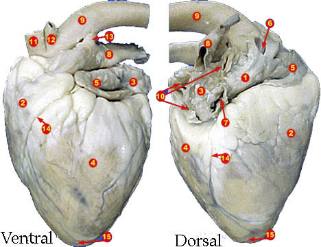
Fig 38.4
Download Printable Version
Lab 38B: Structure of the Heart
PURPOSE:
To review the structural characteristics of the human heart and to examine the major features of a mammalian heart
LEARNING OBJECTIVES:
1. Compare the features of the Human heart with those of other mammalian hearts
PROCEDURE B: Dissection of a Sheep Heart
1. Obtain your sheep heart and rinse it thoroughly, forcing water into the large blood vessels
2. Place your heart in a dissecting pan and examine it carefully. Notice the consistancey of the parietal pericardium. After examining it, remove it by cutting it away. Then locate the visceral pericardium (aka the epicardium) which appears as a thin, transparent layer on the hearts surface. With your scalpel, carefully remove a small portion of the visceral pericardium, exposing the underlying myocardium. You will also note the large amounts of fat lying on the surface of the heart and numerous blood vessels. With the heart ventral-side up, identify the following: atria (right and left), Ventricles (right and left), coronary arteries. Make a quick sketch of the ventral heart and label it in your Lab Notebook
3. Now examine the dorsal surface of the heart. Locate the this walled vessels which enter the right atrium and demonstrate their connection by passing your probe through them. These are the Superior Vena Cava and Inferior Vena Cava. Make a quick sketch of the dorsal side of the heart and label it in your Lab Notebook
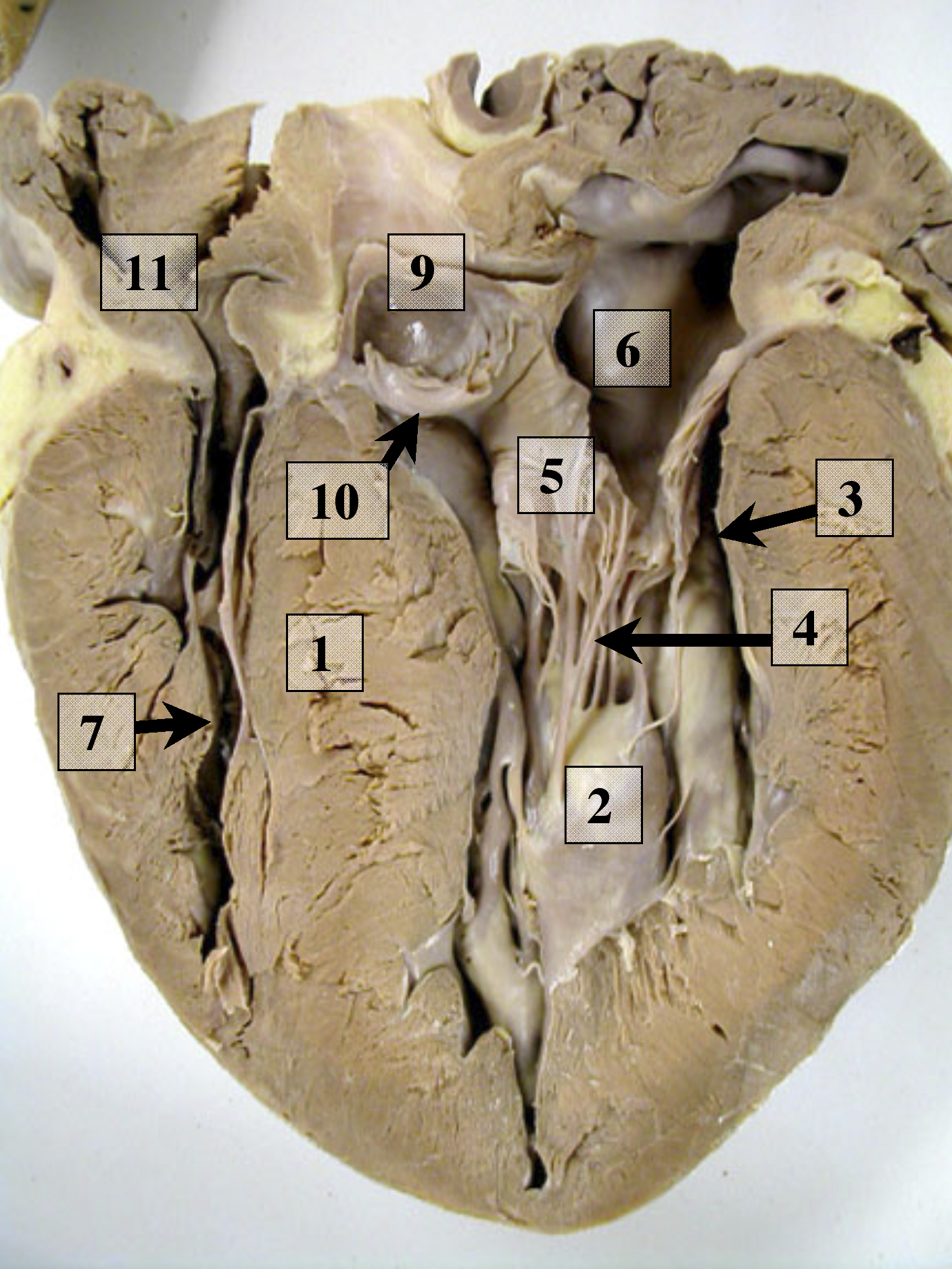
4. Begin your dissection by cutting open the Vena Cava and cutting downward through the lateral edge of the RIGHT atria. Make only a partial cut and then open the heart and examine the tricuspid valve, locating the three "leafs". Also attempt to locate the coronary sinus which is located between the valve and the vena cave (note, it is a small opening). Note that this is where blood feeding the heart returns to circulation for oxygenation.
5. Continue with your cutting by extending it through the wall of the RIGHT ventricle to the apex. Open the ventricle and identify the chordae tendineae and papillary muscles.
6. Locate the opening to the pulmonary trunk and follow it upward out of the ventricle until you find the pulmonary valve. Cut this open and examine the valve. Note how this valve compares to the tricuspid valve.
7. Open the left side of the heart by inserting your scissors into the LEFT atrium and cutting it open. Attempt to find the four openings of the pulmonary veins. Examine the bicuspid valve, and then continue your incision to the apex. Compare the thickness of the walls of the right and left ventricle.
8. Locate the aorta as it leads away from the left ventricle. Compare it in thickness to the other vessels entering and leaving the heart. Cut the aorta open to expose the aortic valve. Examine this valve closely and try to locate the openings of the coronary arteries, which lie just distal of the valve.
9. With your lab partner(s), review the various structures, paying close attention to the vessels associated with the heart and it's four chambers. Cut away a portion of the heart to expose the atria and ventricle from both sides, then make a sketch in your Lab Notebook and label all the specific structures mentioned above.
10. Store your heart as directed.
11. Complete Part B in your Lab Notebook
12. Label Fig. 38.4 and 38.5
13. As you complete the lab, Review the "Lab Objectives" from the handout and write a synopsis of the lab addressing the four objectives.
Fig 38.4
Fig 38.4