Structure of the Respiratory System
Introduction
The respiratory system consists of a group of passages that remove
particles from incoming air and transport it from outside the
body into and out of the lungs. It also includes numerous microscopic
air chambers in which gas exchanges take place between the air
and the blood. The entire process of exchanging gases between
the atmosphere and the body cells is called respiration, and it
involves several events. These include the movement of air in
and out of the lungs - commonly called breathing, or pulmonary
ventilation; the exchange of gases between the air in the lungs
and the blood; the transport of gases by the blood between the
blood and body cells. The utilization of oxygen and production
of carbon dioxide by the cells is called cellular respiration.
Organs of the Respiratory
System
The organs of the respiratory system
include the nose, nasal cavity, sinuses, pharynx, larynx, trachea,
bronchial tree, and lungs. The parts of the respiratory system
can be divided into two sets, or tracts. Those organs outside
the thorax constitute the upper respiratory tract, and
the those within the thorax comprise the lower respiratory
tract.
 |
Nose
The nose is supported internally by bone and cartilage.
Its two nostrils, called the external nares, provide opening
through which air can enter and leave the nasal cavity. These
openings are guarded by numerous internal hairs, which help prevent
the entrance of relatively large particles sometimes carried
in the air.
Nasal Cavity
The nasal cavity, a hollow space behind the nose, is divided
medially into right and left portions by the nasal septum,
which is composed of bone and cartilage.
Nasal conchae
curl out from the lateral walls of the nasal cavity on each side,
dividing the cavity into passageways. They support the mucous
membrane that lines the nasal cavity and help increase its
surface area. The mucous membrane contains pseudostratified
ciliated epithelium that is rich in mucus-secreting goblet
cells.
|
It also includes an extensive network
of
blood vessels, and as air passes over the membrane,
heat leaves the
blood and warms the air. In this way, the temperature
of the incoming
air quickly adjusts to that of the body. In
addition, the incoming
air tends to become moistened by the evaporation
of water from the
mucous lining. The sticky mucus secreted by
the mucous membrane
entraps dust and other small particles entering
with the air.
As the cilia of the epithelial lining move,
a thin
layer of mucus and entrapped particles are
pushed toward the pharynx.
When the mucus reaches the pharynx, it is usually
swallowed. In the
stomach, any microorganisms in the mucus are
likely to be destroyed
by the action of gastric juices.
Paranasal Sinuses
The paranasal sinuses are air-filled
spaces located within (and named from) the
maxillary , frontal, ethmoid, and sphenoid
bones of the skull. These
spaces open into the nasal cavity and are lined
with mucous membranes
that are continuous with the lining of the
nasal cavity.
Although the paranasal sinuses function mainly
to
reduce the weight of the skull, they also serve
as resonant chambers,
which affect the quality of the voice.
Pharynx
The pharynx
(throat) is located behind the oral cavity
and between the nasal
cavity and larynx. It functions as a passageway
for food traveling
from the oral cavity to the esophagus and for
air passing between the
nasal cavity and larynx. It also aids in producing
the sounds of
speech. The pharynx consists of three subdivisions:
nasopharynx (near nasal cavity),
oropharynx (back
of throat, in mouth), and laryngopharynx
(near the
larnyx).
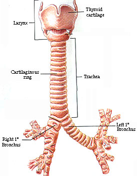
Larynx
The larynx is an
enlargement in the airway at the top of the
trachea and below the
pharynx. It serves as a passageway for air
moving in and out of the
trachea and functions to prevent foreign objects
from entering the
trachea. In addition, it houses the vocal
cords.
 |
The larynx is composed primarily of muscles
and cartilages, which form the framework of the larynx and are
bound together by elastic tissue. The largest of the cartilages
are the thyroid, cricoid, and epiglottic
cartilages.
Inside the larynx, two pair's of horizontal
folds, composed of muscle tissue and connective tissue with a
covering of mucous membrane, extend inward from the lateral walls.
The upper folds are called false vocal cords because they do
not function in the production of sounds. The muscle fibers within
these folds help to close the airway during swallowing.
The lower folds are the true vocal cords.
They contain muscle tissue and elastic fibers and are responsible
for vocal sounds,
|
which are created when air is forced between
the vocal cords, causing vibrations in the
air column above them.
This action generates sound waves, which can
be formed into words by
changing the shape of the pharynx and oral
cavity and by using the
tongue and lips.
The quality of a vocal sound can be altered
by
changing structures within the larynx, pharynx,
and oral cavity.
Thus, the pitch (musical tone) of a sound is
controlled by
contracting or relaxing muscles that alter
the tension on the vocal
folds. Increasing tension produces a higher
pitch, and decreasing
tension causes a lower pitch. The intensity
(loudness) of a sound is
related to the force of the air passing through
the vocal folds.
Louder sound is produced by stronger blasts
of air; softer sound is
produced by weaker blasts of air.
During normal breathing, th vocal cords remain
relaxed, and the opening between them, called
the glottis, appears as a triangular
slit. When food or liquid is swallowed, however,
the glottis is
closed by muscles within the false vocal cords,
and this prevents the
food or liquid from entering the trachea.
The epiglottic cartilage suports a flaplike
structure called the epiglottis. This
structure
usually stands upright and allows air to tenter
the larynx. During
swallowing, however, the larynx is raised,
and the epiglottis is
pressed downward. As a result, the epiglottis
partially covers the
opening into the larynx and helps to prevent
foods and liquids from
entering the air passages.
Trachea
The trachea
(windpipe) is a flexible cylindrical tube about
2.5 cm in diameter
and 12.5 cm in length. It extends downward
in front of the esophagus
and into the throacic cavity, where it splits
into right and left
bronchi.
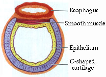 |
The inner wall of the trachea is lined with
a ciliated mucous membrane that contains many goblet cells.
As mentioned before, this membrane continues to remove particles
from the incoming air and to move entrapped particles upward
into the pharynx.
Within the tracehal wall are about twenty
C-shaped peices of hyaline cartilage, arranged one above the
other.
|
The open ends of these incomplete rings
are
directily posteriorly, and the gaps between
their ends are filled
with smooth muscle and connective tissues.
These cartilaginous rings
prevent the trachea from collapsing and blocking
the airway. At the
same time, the soft tissues that complete the
rings in the back allow
the nearby esophagus to expand as food moves
throught it on the way
to the stomach.
Bronchial Tree
The bronchial tree consists of branched
airways leading from the trachea to
the microscope air sacs in the lungs. It begins
with the right and
left primary bronchi,which arise from
the trachia at the level of the fifth
thoracic vertebra.
 |
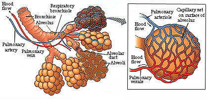
A short distance from its origin, each primary
bronchus divides into secondary bronchi, which in turn
branch again and again into finer and finer tubes. Amoung theses
smaller tubes are some called bronchioles. They continue
to devide, giving rise to very thin tubes called alveolar
ducts. These ducts terminate in groups of microscopic chambers
called alveoli, which are surrounded by capillary nets. |
Although the structure of the bronchus
is
similar to that of the trachea, as finer and
finer branches are given
off, the amount of cartilage in the walls decreases
and finally
disappears in the bronchioles. As cartilage
decreases, however, a
layer of smooth muscle surrounding the tube
becomes more prominenet.
This muscular layer remains even in the smallest
bronchioles, but
only a few muscle fibers occur in the alveolar
ducts.
The branches of the bronchiole tree serve as
air
passages, which continue to remove particles
from the incoming air
and distribute it to the alveoli in all parts
of the lungs. The
alveoli, in turn, provide a large surface area
of thin
squamous epithelial cells through which
gas exchanges can easily occur. During these
exchanges, oxygen diffuses through alveolar
walls and enters the
blood in nearby capillaries, and carbon dioxide
diffuses from the
blood through the walls and enters the alveoli.
It is estimated that there are about 300 million
alveoli in an adult lung and that these spaces
have a total surface
area between 70 and 80 square meters.
Lungs
The lungs are
soft, spongy, cone-shaped organs located in
the thoracic cavity. The
right and left lungs are seperated medially
by the heart and
mediastinum, and they are enclosed by the diaphragm
and thoracic
cage.
Each lung occupies most of the thoracic space
on
its side and is suspended in the cavity by
its attachments, which
include a bronchus and some large blood vessels.
These tubular parts
are connected to the lung on its medial surface.
A layer of serous
membrane, the visceral
pleura, is firmly
attached to the surface
on each lung, and this membrane folds back
to become the
parietal pleura.
The parietal pleura, in turn, forms part of
the mediastinum and lines
the inner wall of the thoracic cavity.
 |
|
The potential space between the visceral and
parietal pleurae is called the pleural cavity, and it
conains a thin film of serous fluid. This fluid lubricated the
adjacent pleural surfaces, reducing friction as they move against
each other during breating. It also helps hold the pleural membranes
together, as explained later.
The right lung is larger than the left one
and is divided into three lobes. The left lung consists of two
lobes.
Each lobe is supplied by a major branch of
the bronchial tree. A lobe also has connections to blood and
lymphatic vessels and is enclosed by connective tissue. Thus,
the substance of a lung includes air passages, alveoli, blood
vessels, connective tissue, lymphatic vessels, and nerves.
|