Animal cells-
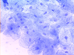
Place a single drop of water on the slide. Use the toothpick to scrape the inner part of your cheek (inside your mouth), removing some cells from your mouth, then rub it into the water drop on the slide. Very carefully add a small drop of dye (methyl blue) to the water, then place the cover slip on top. Examine under the microscope, first under 10x, then under 40x.
Draw a picture following the questions of the cell under high power. Label: cell membrane, cytoplasm, nucleus, the name of the specimen and the magnification.
|
Cheek (10X)
|
Cheek (40X)
|
 |
 |
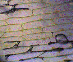
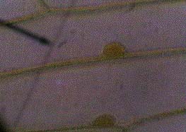
Plant cells- Use a portion of the thin membrane that lines the inside of each "leaf" of an onion. Place it carefully on the slide so that it does not fold upon itself. Place a drop of iodine on the onion instead of methyl blue and cover with the cover slip.. Draw a picture following the questions of the cell under high power. Label: cell wall, cell membrane (if visible), nucleus, cytoplasm
Questions: 1. Why is it important that iodine or methyl blue is added to the specimens? 2. Are the animal and plant cells similar? Explain why or why not. 3. Describe the appearance of the nucleus inside the cells. Does it appear the same in each cell? Is the nucleus in the same position in all cells?